
On 3 May 2026, the World Health Organisation (WHO) issued an alert regarding a possible outbreak of hantavirus on board a Dutch cruise ship travelling from Ushuaia, Argentina, to Cape Verde. It’s not extraordinary for a cruise ship to be afflicted by an infectious disease. However, this is the first instance of a hantavirus outbreak, and circumstances have led public health officials to observe the situation closely. But what do we know about the disease ?
- Hantaviruses are zoonotic viruses found on every continent, transmitted primarily through indirect contact with infected rodents.
- Over 50 hantavirus species have been identified, and an estimated 100,000 human infections occur each year, predominantly in Asia and Europe.
- Hantavirus infections can cause two life-threatening syndromes: haemorrhagic fever with renal syndrome (fatality rate 1–15%) and severe acute cardiopulmonary syndrome with respiratory and cardiac failure (fatality rate 30–60%).
- There are currently no licensed vaccines or treatments.
- A major obstacle to developing hantavirus treatments is the lack of animal models that accurately reproduce human disease: rodents, the natural reservoir, typically carry the virus for life with minimal symptoms, making them poor models for studying how the infection harms people.
- A breakthrough Syrian hamster model that mimics hantavirus pulmonary syndrome in humans has enabled the development of vaccines for Andes, Hantaan, and Puumala hantaviruses, which have now entered Phase I human clinical trials, alongside an antibody-based treatment produced using genetically modified cows.
A zoonotic pathogen found on every continent
The first clinical description of a hantavirus disease dates back to the Korean War (1950–1953), during which more than 3,000 soldiers fell ill. It wasn’t until 1978 that the causative agent was first described by Korean researchers Lee et al., in the lungs of its natural reservoir, the striped field mouse. The virus was named after the Hantaan River, which lies on the border between North and South Korea.
Today, hantaviruses are understood to belong to a large group of viruses that are found on every continent. More than 50 viral species have been identified across the globe, all of which are zoonotic – that is, transmitted by animals. These are mainly wild rodents (rats, voles, field mice) and, more rarely, moles, shrews and bats. Multiple hantaviruses such as Hantaan virus, Seoul virus, Dobrava-Belgrade virus, Puumala virus, Andes virus, and Sin Nombre virus are highly pathogenic to humans. Transmission to humans mainly occurs through indirect contact (saliva, urine, faeces) of asymptomatic infected rodents (referred to as “reservoir” animals) and, more rarely, through direct contact or bites. Human-to-human transmission is rare and appears to involve only the Andes hantavirus – the one that was identified on the cruise ship.
Around 100,000 cases of hantavirus infections are estimated to occur each year, mainly in Asia and Europe. About 90% of all the documented cases worldwide occur in China but Europe totals some 10,000 cases every year in Finland, Germany, Belgium, France, UK, Poland, and the Balkans. In the Americas, the numbers are much lower with a total of eight countries (Argentina, Brazil, Bolivia, Chile, Panama, Paraguay, the USA and Uruguay) reporting only 229 cases and 59 deaths in 2025. However, these numbers are growing every year. Hantaviruses represent a significant and emerging global public health threat, with a devastating effect on human lives and a high mortality rate.
A multi-faceted viral infection
When they affect humans, hantaviruses can cause infections of varying severity. Two types of syndrome may then be observed: haemorrhagic fever with renal syndrome, fatal in 1% to 15% of cases, and severe acute cardiopulmonary syndrome with respiratory and cardiac failure, with a fatality rate of around 30% to 60%. Both syndromes can be caused by the same viral species. However, renal and hepatic syndrome is more commonly found in European and Asian hantaviruses (commonly referred to as “Old World” hantaviruses), while the more dangerous pulmonary syndrome is more commonly observed in American hantaviruses (or “New World” hantaviruses).
Unfortunately, there are currently no licensed treatments or vaccines available to combat hantaviruses. Hantavirus infections are dealt with mostly by managing the symptoms and providing supporting care. The development of vaccines and antivirals remains critical for mitigating hantaviral infections and their impact on public health. However, the mechanisms of pathogenesis are still poorly defined. A better understanding of the virus biology is urgently required, including understanding how infection within rodent carrier populations leads to the emergence of human pathogens and why some of these viruses are able to subvert immune defences and establish high burden disease in humans. These answers will rely on basic research conducted, in part, in animals.
There is still a lot we don’t know about hantaviruses
Medical countermeasures that could held dissipate the threat hantaviruses pose to human populations will require a better understanding of the virus and how it works. This research calls for animal models that faithfully recapitulate the human disease caused by the viral infection. Unfortunately, there are no small animal models that fully recapitulate human haemorrhagic fever with renal syndrome (HFRS) disease. Rodents seem to contract persistent, lifelong infection with minimal symptoms, which allows for a long-term maintenance of the pathogen within the host population, but doesn’t fully reflect what happens in humans.
One of the main reasons we struggle to develop protective or therapeutic options for humans, is the lack of good animal disease models to support the research. Novel antivirals are being tested, some showing promising results, but in vivo testing is often limited to the Andes variant and animal models lacking human-like disease. Without a representative model, it is difficult to investigate potential new drugs.
Several attempts have been made to establish an animal model for hantavirus-like diseases that reflects the human disease. Initial efforts to create a hantavirus nonhuman primate (NHP) disease model were largely unsuccessful. Recently, however, the first hantavirus pulmonary syndrome (HPS) monkey model was described. It emulates HPS human disease but is a logistically complex model, requiring the monkeys to be infected via a deer mouse vector. Rodent models have also been developed more or less successfully. Early pathogenicity studies examined infection in suckling mice and rats with a naturally low immune system, but this often resulted in high lethality. Some immunodeficient mouse models also exhibited some of the human-like symptoms of infection. Despite being limited as pathogenesis (natural disease) models, mice are used extensively in evaluation of vaccine immunogenicity and protective efficacy.
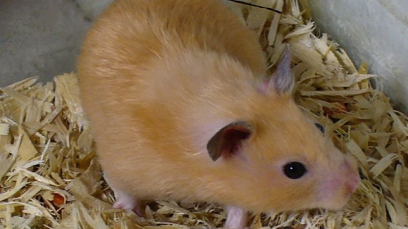
The first real breakthrough was the development of a model for the Andes virus in Syrian hamsters. An immunocompromised version of this model was later adapted to the Sin Nombre virus (SNV). Immunosuppressed Syrian hamsters were infected with various New World hantavirus strains, resulting in an acute disease that mimicked hantavirus pulmonary syndrome in humans. The hamster model has also been used to examine the pathogenesis of hantaviruses and evaluate several vaccines.
Interviewed by Nature, virologist Jay Hooper explained that “a key advance of our team has been developing hamster models of a lethal disease very similar to human hantavirus pulmonary syndrome. This provides a realistic animal model to test vaccines and therapeutics.” Using this model, the team developed vaccines for Andes virus, Hantaan and Puumala hantaviruses that have now reached Phase I human clinical trials. The research team is also developing an antibody-based treatment to combat the Andes virus and three other hantaviruses, that is produced using genetically modified cows.
As animal models get better at reproducing the viral infection in human, the search for new therapeutics and vaccines is likely to become more successful.
Table : Animal models for the evaluation of hantavirus pathogenesis.
|
|
Virus |
Animal model/strain |
% lethality |
Salient features |
|
old world |
HTNV |
ICR suckling mice |
100 |
Only in mice infected within 72 hrs of birth, decreasing thereafter |
|
HTNV |
ICR suckling mice |
97 |
Mice infected 2 to 4 days after birth |
|
|
HTNV |
BALB/c suckling mice |
22.2 |
Mice infected within 24 hrs of birth |
|
|
HTNV |
Striped field mice |
0 |
Antigen, not infectious virus, and persists in lung for 1 yr |
|
|
HTNV |
6–10-week-old Syrian hamsters |
0 |
Asymptomatic infection |
|
|
PUUV |
6–10-week-old Syrian hamsters |
0 |
Asymptomatic infection |
|
|
PUUV |
Suckling and weanling bank voles |
0 |
Asymptomatic, persistent infection |
|
|
PUUV |
Cynomolgus macaques |
0 |
NHP exhibit lethargy, proteinuria, and microhematuria, with histopathological changes in the kidney |
|
|
DOBV |
1–5-day-old NMRI suckling mice |
13–88 |
Viremia, neutralizing antibodies, and elevated levels of NO detected |
|
|
DOBV |
6–8-week-old Syrian hamsters |
0 |
Asymptomatic infection |
|
|
SEOV |
6-day-old Lewis rats |
0 |
Asymptomatic, persistent infection |
|
|
SEOV |
Syrian hamsters (no age specified) |
0 |
Asymptomatic infection |
|
|
SEOV |
70–80-day-old Norway rats |
0 |
Asymptomatic, persistent infection |
|
|
SEOV |
Syrian hamsters (no age specified) |
0 |
Asymptomatic infection |
|
|
new world |
ANDV |
6–8-week-old Syrian hamsters |
Up to 100 |
Recapitulates human disease in incubation time, rapid-progressing respiratory distress, and pathologic findings in the lung |
|
SNV |
6–8-week-old Syrian hamsters |
0 |
Asymptomatic infection, no viremia, and little dissemination Hamster-adapted SNV increases dissemination |
|
|
SNV |
6–8-week-old Syrian hamsters |
100 |
Hamsters immunosuppressed with dexamethasone and cyclophosphamide |
|
|
SNV |
Rhesus macaques |
70 |
Using only deer mouse-passaged SNV |
|
|
SNV |
4–6-week-old deer mice |
0 |
Asymptomatic, persistent infection |
|
|
MAP |
4-week-old Syrian hamsters |
30 |
Recapitulates human disease in incubation time, rapid-progressing respiratory distress, and pathologic findings in the lung |
|
|
PHV |
Cynomolgus macaques/Chimpanzee |
0 |
Mild, transient proteinuria |
Last edited: 21 September 2026 09:47