The 2026 Nobel Prize in Physiology or Medicine was awarded to Karl Deisseroth, Peter Hegemann and Georg Nagel for their work developing optogenetics, a method that shows how nerve cells shape memories, feelings and behaviours in the living brain. Research with frog eggs, hamster cells, rats and mice helped develop the technology, which could contribute to treatments for conditions including blindness, depression, addiction and dementia.
Key takeaways:
- The 2026 Nobel Prize in Physiology or Medicine was awarded to Karl Deisseroth, Peter Hegemann and Georg Nagel for the work that led to optogenetics, a method that shows how nerve cells shape memories, feelings and behaviours in the living brain.
- Optogenetics grew out of research on Chlamydomonas, a light-seeking alga. On the surface of the algea, Hegemann and Nagel discovered channelrhodopsin, a protein that reacts to blue light by creating an electrical impulse. Placed in other cells, it made them light sensitive.
- Researchers can insert the gene for a light-sensing protein into specific nerve cells, then shine light onto them through a thin optic fibre inserted in the brain. This lets them switch individual neural circuits on or off. Deisseroth first did this in the brains of living mice in 2007.
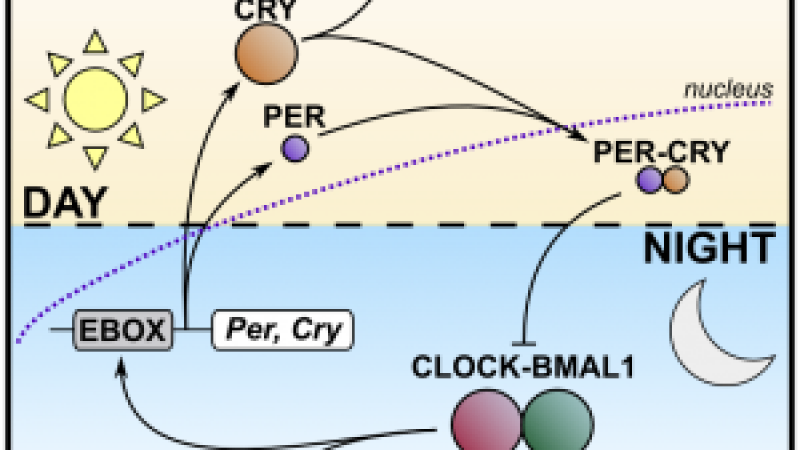
- Optogenetics has helped identify the neurons that control pain, thirst, food intake, reward, attention, and some social behaviours, but also nerve cells that regulate functions beyond the brain, from the circadian rhythm (the body’s daily cycle) to causing a fever when the immune system is activated. It also showed which neurons help form specific fear memories in mice.
- The knowledge gained from optogenetics could contribute to treatments for blindness, depression, addiction and dementia. It has also improved understanding of anxiety, schizophrenia, Alzheimer’s disease and Parkinson’s disease. Researchers are using the method in attempts to restore sight.
For centuries, researchers have tried to understand the brain and how it orchestrates everything from memory to thinking and walking. In the 20th century, they began to investigate which areas of the brain govern different functions, but the methods available could not prove cause and effect. The picture they built of the brain was blurry and disconnected, missing many details. “Optogenetics provides opportunities for mapping the brain in a way that we could once only dream of,” said Per Svenningsson, Chair of the Nobel Committee for Physiology or Medicine.
Born from light-seeking algae
Francis Crick, better known for his part in the discovery of the double helix structure of DNA, was fascinated by the brain and imagined ways to explore its complex networks. Signals in nerve cells are extremely rapid, and Crick speculated that light would be the ideal tool to match that speed. He predicted that if nerve cells could be made to react to light, scientists might be able to control neural activity with precise timing and location. But even Crick admitted that his idea sounded far-fetched.
In the 1990s, however, one researcher’s curiosity began turning Crick’s vision into reality. “The solution didn’t come from studying the brain – it came from a pond, with the humble green algae as an unlikely hero,” Abdel El Manira, a neuroscientist at the Karolinska Institute and a member of the Nobel Committee, told the Scientist. The alga Chlamydomonas was famous for its ability to swim towards light. Peter Hegemann, at the Max Planck Institute for Biochemistry in Martinsried, asked a simple question: how can this single-celled alga react so dynamically to light?
In the early 2000s, Hegemann and Georg Nagel discovered channelrhodopsin, a protein on the surface of the algal cell that reacts to blue light. When light hits it, the protein opens and electrically charged particles (ions) flow into the cell, creating an electrical impulse. The protein was hard to study, because it became damaged and unstable when researchers tried to isolate it. However, scientists mapped the DNA of Chlamydomonas and found the genes behind the protein. This opened up a whole new avenue of research, because the light-capturing protein could now be inserted into any cell they chose.
Hegemann and Nagel introduced the protein into frog egg cells, then into embryonic human kidney cells and hamster kidney cells. Whichever cell they used, it became light sensitive and produced an electrical signal when exposed to light. “Nagel and Hegemann realized exactly what they had found,” said El Manira. “They had just discovered the switch neuroscientists had long dreamed of.” The researchers published their results in 2003. Not long after, a young scientist at Stanford University took up the baton.
A light switch to the brain
Karl Deisseroth wrote to Nagel to request the DNA of the light-sensing protein, hoping to use it to trigger impulses in nerve cells. He first introduced the gene into rat nerve cells grown in petri dishes. By illuminating the cells with blue light, he triggered a nerve signal that was passed on to other neurons.
Deisseroth’s research group published its discovery in 2005. It was a decisive milestone, but there was still a long journey ahead before reaching Deisseroth’s goal: controlling the activity of nerve cells in live animals. He began numerous collaborations, including with Peter Hegemann and Georg Nagel. With Deisseroth’s lab at the forefront, researchers soon found more proteins that could turn nerve cells on and off, activated by different wavelengths of light.
In 2006, this new method was given the name optogenetics. One year later, Deisseroth successfully activated nerve cells in the brains of living mice. His group introduced the gene for the light-sensing protein into a specific group of nerve cells in the motor cortex, the area of the brain that governs movement. A small hole was drilled in the animal’s skull so that a thin optic fibre could shine light onto these neurons. Illuminating this group of cells allowed the researchers to control the movements of the animal’s whiskers.
The same year, working with other researchers, Deisseroth used optogenetics to wake sleeping mice in a controlled manner, helping to confirm which neurons control wakefulness. Another famous piece of work from the early days of optogenetics was published in 2012. Together with Susumu Tonegawa (winner of the 1987 Nobel Prize for discoveries in immunology), Deisseroth studied a pattern of neural activity that formed memories when mice felt fear. When the researchers later reactivated these nerve cells using optogenetics, the mice showed signs of fear even though they were in no danger. This was the first demonstration of particular neurons helping to create specific memories.
A new era in understanding the brain
Crick’s vision had become reality. Optogenetics finally provided the long-sought super fast switch for neuronal activity. An adult human brain has around 90 billion nerve cells, each forming thousands of connections with other nerve cells. Using light, researchers can now switch individual neural circuits on or off in the living brain. They can bring memories to life, create feelings, drive behaviours and study the types of neurons involved in various psychiatric and neurological disorders.
“The impact of optogenetics extends far beyond understanding the healthy brain,” said El Manira. Optogenetics is now used in laboratories around the world to reveal the brain’s mysteries. Neuroscience has moved from passively recording brain activity to actively controlling it, illuminating how the brain encodes cognition, emotion and behaviour. Since its discovery, optogenetics has helped researchers pinpoint exactly which neurons control different brain functions, including pain, thirst, food intake, reward, attention, and some social behaviours. Optogenetics studies of complex behaviours, such as how mice care for their young, have shown that the different components of a behaviour can be controlled by separate neural circuits. One circuit governs, for example, how mice gather their young in a nest; another regulates how they groom them. Scientists have also identified nerve cells that regulate functions beyond the brain, from the circadian rhythm (the body’s daily cycle) to causing a fever when the immune system is activated.
The hope that this knowledge will lead to new medical treatments has begun to come to fruition. Optogenetics has provided greater understanding of the distinguishing features of psychiatric and neurological disorders such as depression, addiction, anxiety, schizophrenia, Alzheimer’s disease and Parkinson’s disease. Researchers have also taken the first steps towards using optogenetics as a medical treatment, restoring sight in people with visual impairment.
FAQ:
What is optogenetics?
Optogenetics is a method that uses light to switch nerve cells on or off. It can be used in a live animal. It has helped show how nerve cells shape memories, feelings and behaviours.
Who won the 2026 Nobel Prize in Physiology or Medicine?
Karl Deisseroth, Peter Hegemann and Georg Nagel were awarded the prize for their work that led to optogenetics.
How was optogenetics discovered?
Peter Hegemann and Georg Nagel studied how the alga Chlamydomonas, swims towards light. They discovered channelrhodopsin, a protein on the surface of the alga’s cell that reacts to blue light. When light hits it, the protein opens and electrically charged particles (ions) flow into the cell, creating an electrical impulse.
What is channelrhodopsin?
Channelrhodopsin is a light-sensitive protein. When light hits it, the protein opens and electrically charged particles (ions) flow into the cell, creating an electrical impulse. When it was placed on the surface of frog egg cells, embryonic human kidney cells and hamster kidney cells, those cells became light sensitive.
How does optogenetics work in the brain?
Researchers insert the gene for a light-sensing protein into specific nerve cells. They then shine light onto those cells through a thin optic fiber that is inserted into the brain to the zone of interest. In 2007, Deisseroth’s team were able to control the movements of mice’s whiskers using optogenetics.
Why is using light so important to control nerve cells?
Signals in nerve cells are extremely rapid. Francis Crick speculated that light would be the ideal tool to match that speed. He predicted that scientists might be able to control neural activity with precise timing and in precise locations. Optogenetics finally provided the long-sought switch for neuronal activity, with millisecond precision.
What animals were used to develop optogenetics?
Frog eggs, hamster cells, rats and mice helped develop optogenetics.
What has optogenetics taught us about the brain?
Optogenetics has helped researchers pinpoint which neurons control pain, thirst, food intake, reward, attention, and social behaviours such as caring for the young. In mice, it notably showed that separate neural circuits can control different parts of a behaviour, such as gathering young in a nest and grooming them. It has also identified nerve cells that regulate the circadian rhythm and cause a fever when the immune system is activated.
Can optogenetics bring back memories?
In a 2012 study, Deisseroth and Susumu Tonegawa reactivated the nerve cells that formed a fear memory in mice using optogenetics. The mice showed signs of fear even though they were in no danger. This was the first demonstration of which neurons help create specific memories.
Can optogenetics treat disease?
Not yet as a routine treatment. The technology could contribute to treatments for conditions including blindness, depression, addiction and dementia. Optogenetics has notably provided a greater understanding of the distinguishing features of psychiatric and neurological disorders such as depression, anxiety, schizophrenia, Alzheimer’s disease and Parkinson’s disease.
REFERENCES:
Harz, H., Nonnengasser, C., Hegemann, P. The photoreceptor current of the green alga Chlamydomonas. Philos. Trans. R. Soc. Lond. Ser. B: Biol. Sci. 1992:338:39–52.
Nagel G, Ollig D, Fuhrmann M, Kateriya S, Musti AM, Bamberg E, Hegemann P. Channelrhodopsin-1: a light-gated proton channel in green algae. Science. 2002:296:2395–2398.
Nagel G, Szellas T, Huhn W, Kateriya S, Adeishvili N, Berthold P, Ollig D, Hegemann P, Bamberg E. Channelrhodopsin-2, a directly light-gated cation selective membrane channel. Proc. Natl. Acad. Sci. U. S. A. 2003:100:13940–13945.
Boyden ES, Zhang F, Bamberg E, Nagel G, Deisseroth K. Millisecond-timescale, genetically targeted optical control of neural activity. Nat. Neurosci. 2005:8:1263–1268.
Aravanis AM, Wang LP, Zhang F, Meltzer LA, Mogri MZ, Schneider MB, Deisseroth K. An optical neural interface: in vivo control of rodent motor cortex with integrated fiberoptic and optogenetic technology. J Neural Eng. 2007:4:S143–156.
Gholami Pourbadie H, Sayyah M. Optogenetics: Control of brain using light. Iran Biomed J. 2018;22(1):4-5.
Hegemann P, et al. Algal sensory photoreceptors. Phycol J.2001;37(5):668-676.
Liu X, et al. Optogenetic stimulation of a hippocampal engram activates fear memory recall. Nature. 2012;484(7394):381-385.
Emiliani V, et al. Optogenetics for light control of biological systems. Nat Rev Methods Primers. 2022;2:55.
Zhou Y, et al. Optogenetics in medicine: Innovations and therapeutic applications. Curr Opin Biotechnol. 2025;92:103262.
Last edited: 20 October 2026 09:07