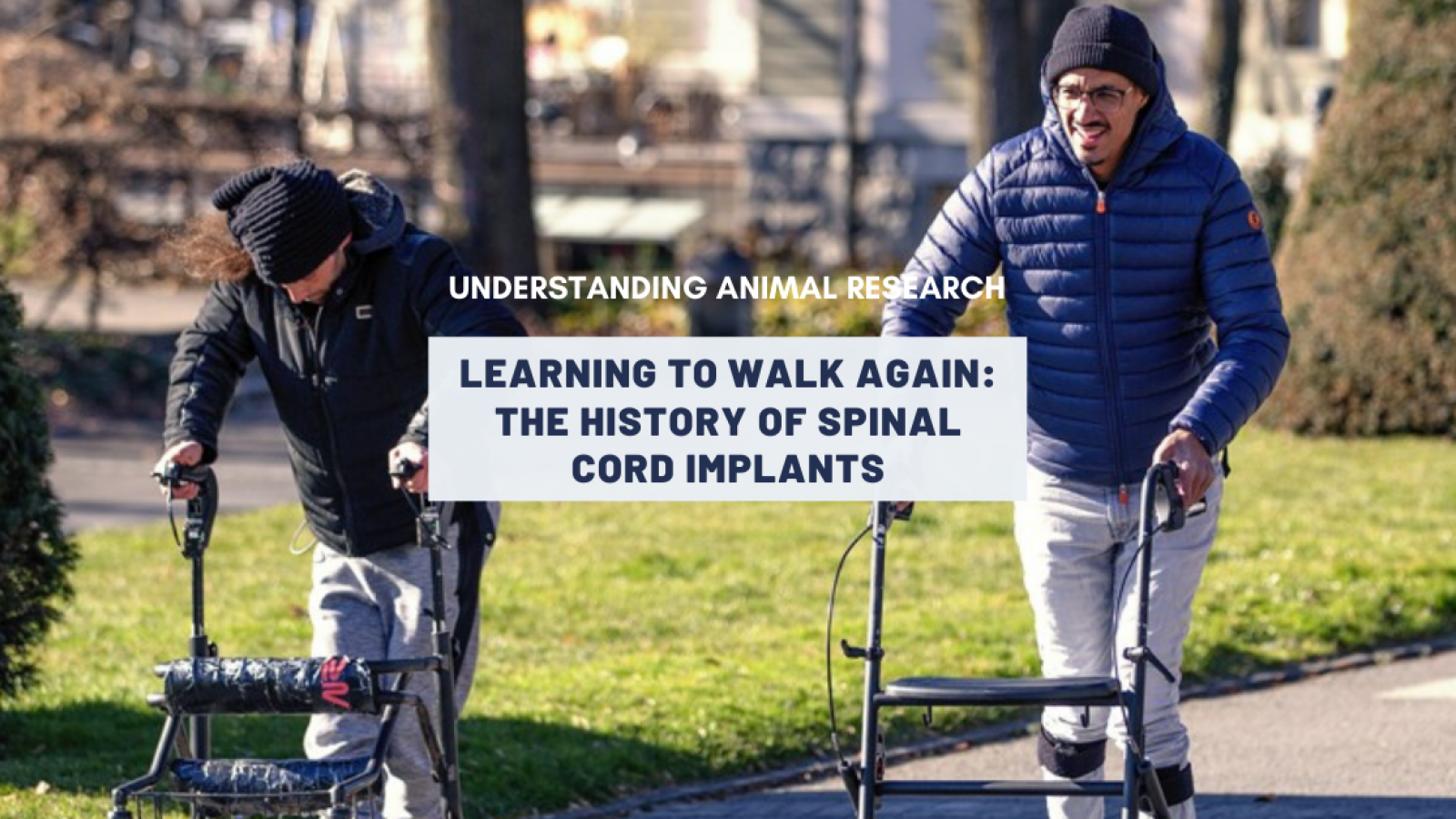
Five years ago, a motor bike accident completely severed Michel Roccati’s spinal cord. He was left with no feeling at all in his legs and paralysed from the waist down. Last month, he was able walk again thanks to an electrical implant surgically attached to his spine.
This is a major step at improving quality of life for patients with spinal cord injuries. It is the result of three decades of research, including vital studies in rabbits, rats, sheep, cats and monkeys.
When the spinal cord is severed, the electrical information from the brain cannot reach parts of the body. The electrical flow is disrupted even though the neurones below the injury remain intact. For decades, researchers have focused on repairing those connections but it was a technique originally developed for something quite different that led to the recent breakthrough.
Spinal cord stimulation delivered by electrical implants was first used in 1967 to treat chronic pain. A mild electrical stimulation of the nerves along the spinal column modified nerve activity and minimised the sensation of pain reaching the brain. As the years went by, newer designs provided more precise control of the electrical field and increasingly sophisticated devices until Susan Harkema, a neurophysiologist at the University of Louisville in Kentucky, speculated that a similar approach might allow people with spinal-cord injuries to stand and walk by replacing the signals that once came from the brain and bridging the gap created by injury. And it did.
In 2009, a patient of Harkema’s, Rob Summers, was implanted with a strip of tiny electrodes in his spine, and gradually regained toe wiggle function. Findings from Harkema’s group and other laboratories suggested that some connections in the spinal cord do in fact remain intact, even for people with the most severe damage. Electrical stimulation seemed to help amplify the messages sent across the injuries, and re-establish these links.
A history of animal research
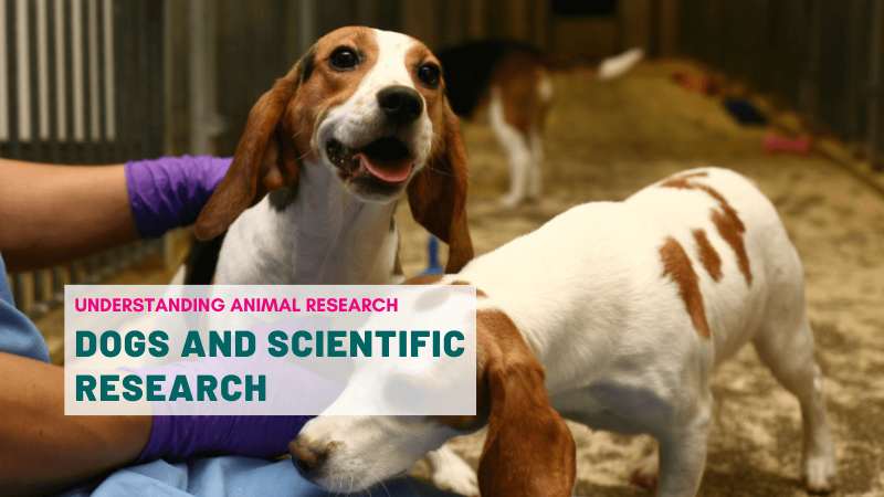
The path to Summers’s toe wiggle began a long time ago, with rabbits. Because spinal cord injury affects the whole body, pre-clinical research is currently dominated by the use of animals to model injuries in humans, and replacing animals for this type of research is challenging at present.
The first experimental animal model for spinal cord injury was developed by the German pathologist Hans Schmaus in 1890. He studied the effect of concussion on a rabbit’s spinal cord. The injury was induced by tying a wooden board to a rabbit's back and performing a blow trauma to the board. Over the years, various mechanisms for inducing spinal cord injury were tested from weight drop methods modelled in dogs to paraffin injections to produce compression injury in cats. In the 1950s, Tarlov developed a hydraulic compression device to produce both acute and chronic compression injuries with quantitative degenerative lesions in dogs. Various animal species have also been used, including rhesus macaques, Canadian geese, Peking ducks, guinea pigs, rats, mice and sheep to help understand the mechanism and the pathophysiology of injury, and to develop reliable treatment strategies.
Today, contusion and compression methods are the most common patterns of injury to best simulate the biomechanics and neuropathology of human injury, whereas transection models are valuable to study anatomic regeneration. Rodents are the most common and probably best-suited species for preliminary spinal cord injury studies. However spinal injury is common in dogs, particularly dachshunds, and many dog owners are glad to let research be performed on their pets, which might otherwise have to be put down.
Because these types of experiments on animals can involve significant injury and have the potential to cause significant levels of suffering, they are heavily regulated by the UK Home Office. A great deal is done to minimise animal suffering and to apply the 3Rs. Recommendations are available to refine the methods and reduce the number of animals used, which are highly effective ways to reduce suffering and improve scientific quality.
From cats on treadmills to humans
All the previous work in animals led to Reggie Edgerton, a physiologist at the University of California, to start working in the 1970s with cats that had had their spinal cord severed. Suspended over a treadmill, the animals could be trained to walk again by simply guiding their legs in a step-like motion, even without any input from the brain. The spinal circuitry propelling them forward. This circuitry is called a ‘central pattern generator’.
In 1993, Harkema joined Edgerton’s lab and began doing similar experiments in humans who had spinal cord injuries. It worked to some extent but Harkema and Edgerton wanted to see a bigger effect. Electrical stimulators seemed like a good option. Indeed, in 2002, researchers in Arizona reported that a 43-year-old man with an incomplete spinal injury was able to walk with coordinated locomotion patterns, much like the cats, when he was placed over a moving treadmill and had his spine stimulated.
In December 2009, Harkema’s team fitted Rob Summers with an epidural stimulator. They placed a 16-electrode array in the space between his vertebrae and his spinal cord. Within three days he could stand with assistance and then walk over the treadmill. Six months later he regained voluntary toe movement. This was an incredible achievement. Somehow, the brain command was reaching the lower body. Soon, other research teams were trying the approach in humans, and looking to see whether it could allow them to take steps off the treadmill.
Grégoire Courtine, a neuroscientist at the Swiss Federal Institute of Technology in Lausanne (EPFL) and an old colleague of Harkema’s, was studying epidural stimulation in rodents, and eventually in rhesus macaques. By 2015, he felt ready to test the technology in humans. His team used the same electrical stimulator Harkema used, but tweaked the software so that the device delivered patterns of stimulation timed to coincide with the act of walking. He was trying to activate the spinal cord, as the brain tried to do.
After years of training and stimulations, the patients with incomplete spinal injuries managed assisted walking.
From partially walking to fully walking
Earlier this year, Courtine brought new hope to patients with complete spinal cord injury. He used his tailored, brain mimicking, stimulating implant to help three of his patientswalk, swim, work the pedals of a bicycle and even paddle canoes.
Courtine’s group used magnetic resonance imaging and computed tomography to map the neurons in the spinal cords, and create a predictive model of the average spinal cord. This helped indicate where precisely to place the implant’s electrodes. The researchers then fine-tuned the electrical current to each individual. Once the implant was in place, each person could control the pattern of electrical stimulation, using buttons and a tablet to raise or lower each leg.
All three participants recovered some level of movement within one day of activating the implant, including being able to walk on a treadmill while their weight was supported. After months of training, the participants were also able to take part in activities such as pedalling a bicycle and performing squats, using the device to guide their muscles through pre-programmed movements.
Courtine says that his group hopes to simplify the technology so that a user can control it through a smartphone. The team has received approval from the US Food and Drug Administration to test the system in a US-based clinical trial with more people. Courtine says the researchers plan to focus on people with recent injuries, because tests in rodents have shown that animals tend to recover more motion when the stimulation begins immediately after their injury.
Research is still ongoing
Still, researchers are cautious. Although the stimulators are approved by the US Food and Drug Administration to treat chronic pain, they do occasionally cause side effects such as shock, burns or nerve damage. Stimulators have been named in almost 80,000 injury reports since 2008 — more than for any other medical device, apart from insulin pumps and metal hip replacements.
Alternative high-tech approaches to regain movement, such as brain–machine interfaces which are potentially less risky, are also being developed. In 2016, a man was able to move his arm by using an interface that could decode his thoughts and electrically stimulate his own muscles. Such a ‘neural bypass’ had been previously used successfully in monkeys. Similarly, after work in rats and monkeys, a man was implanted with electrodes that could read brain activity so he could control his legs. He performed the symbolic kick-off for the World Cup in Brazil in 2014. In 2016, Australian scientists designed in sheep a "bionic spine", a tiny 3cm long device that could enable paralysed patients to walk again by allowing them to control bionic limbs with the power of subconscious thought.
Whether it is stem cell research, combination therapies, exoskeletons, or implants, there are many ways research is looking to help the several million people worldwide, and some 2,000 people a year in the UK, who suffer traumatic spinal cord injury. In almost all cases, the research requires work with animals.
Last edited: 4 April 2022 16:42