Ovarian cancer is the deadliest gynecologic cancer. It kills nearly 4,200 women in the UK every year. Although Ovarian cancer survival is improving and has almost doubled in the last 40 years in the UK, it still kills 6 in 10 women in the first 5 years of the disease.
One of the reasons for such a low survival rate is that it tends to be diagnosed at a relatively late stage. Although surgery works really well to block the cancer when it is still confined to the ovaries and fallopian tubes, once the cancer has spread beyond there and cancer cells are lodged in different parts of the body, even chemotherapy does not prevent the disease.
“The drugs that are used are often 30 or 40 years old and there's been little improvement in the field for quite some time. A few targeted therapies look really promising but probably only for a subset of patients. So there is really an unmet need within ovarian cancer and real room for refinement. There's a lot we can learn about the biology of the cancer that could translate to future therapies,” explains Dr Patrick Caswell, Senior Lecturer at Wellcome Trust Centre for Cell-Matrix Research at The University of Manchester.
Why is ovarian cancer different?
The metastatic spread of ovarian cancer is really the most lethal aspect of the disease. Once the cancerous cells start colonising different places in the body, resistance to therapy appears and the real problems start. But unlike other cancers that spread through the bloodstream, ovarian cancer cells are generally confined within the peritoneal cavity* around the organs which can make them difficult to access.
Ovarian cancer cells also mutate at a high rate. They seem to be able to adapt quite quickly and become resistant to chemotherapy.
Targeting the tumour micro-environment
To try to overcome the adaptability of ovarian cancer cells, researchers have tried to focus on the cells that surround and succour the cancer cells, the cells that make up the tumour micro-environment. The cells in the micro-environment around tumours provide information about where cells are in tissues and can instruct cells on where to grow and to divide to avoid cell death. Cells such as fibroblasts also give structural support and so modifying or disrupting this supportive micro-environment has become a key target for new therapies.
Influencing these cells could give clinicians tools to prevent cancer cell growth. Although tumour microenvironment cells are associated with the cancer and often shape the conditions in which the tumours grow, they are more stable and less likely to mutate which makes them good targets for drugs and other therapeutic interventions.
Collecting tumour micro-environments from patients
Researchers work in collaboration with clinicians and gynaecological surgeons to collect patient-derived living cells and tissues to study them. They extract the different cell-types and grow them in culture.
It is relatively easy to separate cancerous cells from microenvironment cells and grow them separately. Scientists can then grow them back together and observe how their behaviours change and how they affect one another. It is also possible to add other cells, like fatty cells that provide a source of energy for cancer cells, to see how they fit in the picture. This is called an organo-typic model and provides a 3D representation in the laboratory of what is happening in the human body. It helps researchers reconstruct, bit by bit, what is going on within the patients.
But looking at these cells in the lab isn’t enough. The human cells help understand which proteins are important and the mechanisms at play. But this needs to hold true in whole organisms before conclusions can be made in humans and therapies contemplated.
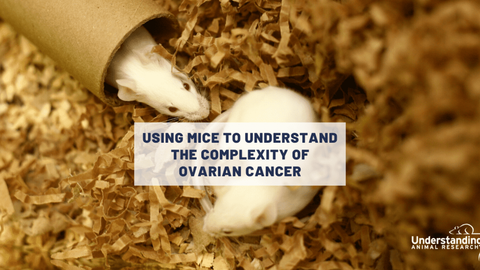
Animal models are crucial to understanding the tumour microenvironment
Over the past 5 to 10 years, there’s been a growing realisation that the tumor micro-environment is a really complex structure. All sorts of different cell types, with different growth factors and immune signals, come into play and communicate with the cancerous cells. In fact, the cell-based approach is a very reductionist way of looking at things. It doesn’t help grasp the complexity of the interactions.
“Although technically, it could be possible to isolate each compound involved, for now it is still impossible to reassemble everything to mimic the living tissue as a whole. Fundamentally it’s difficult to recreate the interactions between the cancer cells, the tumor micro-environment and then the immune system. This is where animal models are important,” explains Patrick Caswell.
Mice genetically engineered to have the mutated genes that can frequently be found in human ovarian cancer cells go on to develop a disease that deeply resembles ovarian cancer in humans. Patrick Caswell uses these mice to look at the tumor microenvironment in a whole organism and see how affecting that microenvironment influences the cancer’s ability to grow. For example, he injects tumour microenvironment cells into the peritoneal space of healthy mice to see what happens.
Animal models can also help understand how drugs can affect the ability of microenvironment cells to make the right niche for the cancer cells to grow – or not.
New animal models could be the boost the field needs
In the past, mouse models for ovarian cancers have proved to be really difficult to create. It’s only been 8 or 9 years that these mouse models have started to improve and bring new information to the table.
“I guess the lack of funding with regards to other cancers, has meant that the level of information needed to understand the biology and create these models wasn’t there,” explains Patrick Caswell. “For example, we now know that high grade serious ovarian cancer has a mutation frequency of somewhere above 96% in a tumor suppressor gene called P53. In early models that wasn't known. So that really important information wasn't included in the mice models. By gradually increasing the knowledge of the biology, we are improving the animal models we have at hand, and hence inching closer to better fighting the disease.”
In the past five years, he can point to two or three papers that have shaken the field. And these findings were only made possible because we had shown beforehand that the microenvironment within these animal models really closely matches what we find in patients.
“That's probably one of the biggest steps we've made very recently in the field,” Patrick Caswell adds. “My personal feeling is that the use of animal models probably hasn't changed in terms of the volume, but we have become more savvy about the types of model we use. Not only have we become better at taking cells from patients, which means that we are not relying strains that were harvested some 20 years back, and growing them in a much more realistic environment, but we now have mouse models that more accurately reflect the genetic changes that we see in the cancer cells and really do closely recapitulate the characteristics of the disease. This will be a real game changer.”
* The peritoneal cavity is between the parietal peritoneum membrane (the peritoneum that surrounds the abdominal wall) and visceral peritoneum (the peritoneum that surrounds the internal organs) https://en.wikipedia.org/wiki/Peritoneal_cavity
Last edited: 21 June 2023 15:24